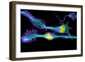
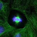
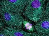
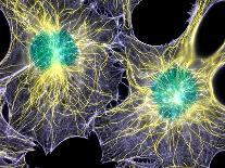
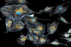
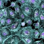
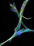
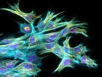
Nerve cancer cells Immunofluorescence light micrograph of cultured cells from a neuroblastoma a type of nerve tissue tumour Fluorescent dyes have been used to highlight proteins in the cell nuclei yellow and cytoskeletal protein filaments tubulin green forming microtubules and actin blue The cells have been stimulated to undergo neural differentiation and have changed from a spherical shape to form long branching extensions called neurites green blue and purple These are designed to extend outwards and connect to other neurons A neuroblastoma is a malignant cancerous tumour most commonly found in children that derives from primitive nerve cells in a kidneys adrenal gland Immunofluorescence uses antibodies to attach fluorescent dyes to tissues and molecules in a cell Magnification x when printed cm wide