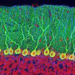
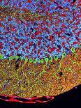
Retina cells. Fluorescent light micrograph of cells in the retina, the lightsensitive membrane that lines the back of the eyeball. A blood vessel runs from left to right, and numerous other branches are seen. Astrocyte glial cells are green. The glial cells provide structural support to the nerve cells that send visual signals to the brain. The tissue has been tagged with fluorescent markers specific to certain proteins. The greenmarks the glial fibrillary acidic protein (GFAP) found in glial cells. Blue marks plateletendothelial cell adhesion molecule1 (PECAM1) found in blood vessels, and red is the structural protein actin.