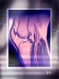
Knee joint, coloured magnetic resonance imaging (MRI) scan. In this side view, the front of the knee is at left. The patella (kneecap) is at far left. The knee is the largest joint in the body. It is formed from the articulation of the femur (thigh bone, upper centre) and the tibia (shin bone, lower centre). Behind these bones are supporting muscles and tendons. MRI scanners use a powerful magnet to align hydrogen atoms in body tissues. A radio frequency pulse is used to knock these out of alignment. The energy that the atoms emit as they realign themselves with the field is used to build up an image of tissues.