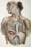
Kidney stone in ureter. Coloured urogram (Xray) of a patients abdomen showing a blocked ureter due to a kidney stone (calculus). The ureters join the kidneys (blue, upper left & right) to the bladder (bottom centre, white). The renal pelvis of each kidney that collects urine is pink. A dye (blue) highlights the long ureters. The ureter at right is wide, and the renal pelvis enlarged, due to urine pooling in them unable to pass into the bladder. This is due to a kidney stone obstructing the lower part of the ureter. Kidney stones form when urine salts harden. Blockage of the ureter can cause pain and kidney damage. The stone can be surgically removed or broken up by lithotripsy.