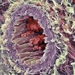
Inner ear sensory cells. Coloured scanning electron micrograph (SEM) of bundles of cilia (hair cells, blue) situated in the macula utriculi within the labyrinth of the human inner ear. Each bundle comprises many stereocilia and one long kinocilium. They detect directional movement, such as the tilting of the head. The cilia are immersed in a fluid, called the endolymph. Currents in this fluid displace the position of the cilia, resulting in the production of pulses, which are sent to the brain along the vestibular nerve. The surrounding support cells are covered in projections called microvilli (brown). Two otoliths (purple) are also seen. These crystals also help sense balance.