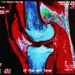
Knee joint. Coloured Magnetic Resonance Imaging (MRI) scan of a sagittal section through a human knee joint. Two bones (coloured blue) meet at the knee, forming a hingejoint. At top is the femur (thighbone), which articulates with the tibia (shinbone) at bottom. The patella or kneecap (red, located at upper left) is a protective bone at the front of the knee held in position by muscles and tendons. Two discs of protective cartilage cover the surfaces of the femur and tibia to reduce friction between these bones. The round bottom surface of the femur forms a narrow ridge that moves through a groove in the tibia. This allows hinge movement, with slight rotation.